Cell Structure :: Ideas about cell structure have changed considerably over the years. Early biologists saw cells as simple membranous sacs containing fluid and a few floating particles. Today’s biologists know that cells are infinitely more complex than this.

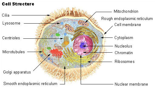
There are many different types, sizes, and shapes of cells in the body. For descriptive purposes, the concept of a “generalized cell” is introduced. It includes features from all cell types. A cell consists of three parts: the cell membrane, the nucleus, and, between the two, the cytoplasm. Within the cytoplasm lie intricate arrangements of fine fibers and hundreds or even thousands of miniscule but distinct structures called organelles.
Cell membrane
Every cell in the body is enclosed by a cell (Plasma) membrane. The cell membrane separates the material outside the cell, extracellular, from the material inside the cell, intracellular. It maintains the integrity of a cell and controls passage of materials into and out of the cell. All materials within a cell must have access to the cell membrane (the cell’s boundary) for the needed exchange.
The cell membrane is a double layer of phospholipid molecules. Proteins in the cell membrane provide structural support, form channels for passage of materials, act as receptor sites, function as carrier molecules, and provide identification markers.
Nucleus and Nucleolus
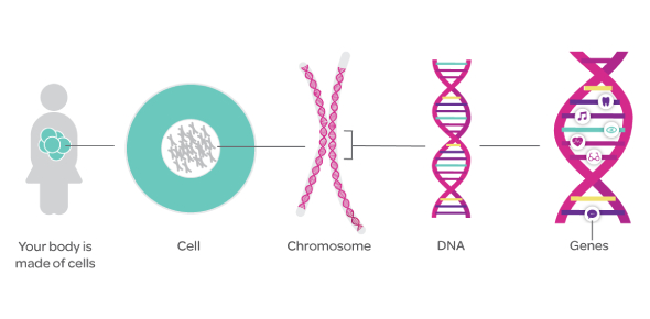
The nucleus, formed by a nuclear membrane around a fluid nucleoplasm, is the control center of the cell. Threads of chromatin in the nucleus contain deoxyribonucleic acid (DNA), the genetic material of the cell. The nucleolus is a dense region of ribonucleic acid (RNA) in the nucleus and is the site of ribosome formation. The nucleus determines how the cell will function, as well as the basic structure of that cell.
Cytoplasm
The cytoplasm is the gel-like fluid inside the cell. It is the medium for chemical reaction. It provides a platform upon which other organelles can operate within the cell. All of the functions for cell expansion, growth and replication are carried out in the cytoplasm of a cell. Within the cytoplasm, materials move by diffusion, a physical process that can work only for short distances.
Cytoplasmic organelles
Cytoplasmic organelles are “little organs” that are suspended in the cytoplasm of the cell. Each type of organelle has a definite structure and a specific role in the function of the cell. Examples of cytoplasmic organelles are mitochondrion, ribosomes, endoplasmic reticulum, golgi apparatus, and lysosomes.
Cell Function
The structural and functional characteristics of different types of cells are determined by the nature of the proteins present. Cells of various types have different functions because cell structure and function are closely related. It is apparent that a cell that is very thin is not well suited for a protective function. Bone cells do not have an appropriate structure for nerve impulse conduction. Just as there are many cell types, there are varied cell functions. The generalized cell functions include movement of substances across the cell membrane, cell division to make new cells, and protein synthesis.
Movement of substances across the cell membrane
The survival of the cell depends on maintaining the difference between extracellular and intracellular material. Mechanisms of movement across the cell membrane include simple diffusion, osmosis, filtration, active transport, endocytosis, and exocytosis.
Simple diffusion is the movement of particles (solutes) from a region of higher solute concentration to a region of lower solute concentration. Osmosis is the diffusion of solvent or water molecules through a selectively permeable membrane. Filtration utilizes pressure to push substances through a membrane. Active transport moves substances against a concentration gradient from a region of lower concentration to a region of higher concentration. It requires a carrier molecule and uses energy. Endocytosis refers to the formation of vesicles to transfer particles and droplets from outside to inside the cell. Secretory vesicles are moved from the inside to the outside of the cell by exocytosis.
Cell division

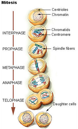
Cell division is the process by which new cells are formed for growth, repair, and replacement in the body. This process includes division of the nuclear material and division of the cytoplasm. All cells in the body (somatic cells), except those that give rise to the eggs and sperm (gametes), reproduce by mitosis. Egg and sperm cells are produced by a special type of nuclear division called meiosis in which the number of chromosomes is halved. Division of the cytoplasm is called cytokinesis.
Somatic cells reproduce by mitosis, which results in two cells identical to the one parent cell. Interphase is the period between successive cell divisions. It is the longest part of the cell cycle. The successive stages of mitosis are prophase, metaphase, anaphase, and telophase. Cytokinesis, division of the cytoplasm, occurs during telophase.
Meiosis is a special type of cell division that occurs in the production of the gametes, or eggs and sperm. These cells have only 23 chromosomes, one-half the number found in somatic cells, so that when fertilization takes place the resulting cell will again have 46 chromosomes, 23 from the egg and 23 from the sperm.
Heredity, Genes, and DNA :: Perhaps the most fundamental property of all living things is the ability to reproduce. All organisms inherit the genetic information specifying their structure and function from their parents. Likewise, all cells arise from preexisting cells, so the genetic material must be replicated and passed from parent to progeny cell at each cell division. How genetic information is replicated and transmitted from cell to cell and organism to organism thus represents a question that is central to all of biology. Consequently, elucidation of the mechanisms of genetic transmission and identification of the genetic material as DNA were discoveries that formed the foundation of our current understanding of biology at the molecular level.
Genes and Chromosomes
The classical principles of genetics were deduced by Gregor Mendel in 1865, on the basis of the results of breeding experiments with peas. Mendel studied the inheritance of a number of well-defined traits, such as seed color, and was able to deduce general rules for their transmission. In all cases, he could correctly interpret the observed patterns of inheritance by assuming that each trait is determined by a pair of inherited factors, which are now called genes. One gene copy (called an allele) specifying each trait is inherited from each parent. For example, breeding two strains of peas—one having yellow seeds, and the other green seeds—yields the following results The parental strains each have two identical copies of the gene specifying yellow (Y) or green (y) seeds, respectively. The progeny plants are therefore hybrids, having inherited one gene for yellow seeds (Y) and one for green seeds (y). All these progeny plants (the first filial, or F1, generation) have yellow seeds, so yellow (Y) is said to be dominant and green (y) recessive. The genotype (genetic composition) of the F1 peas is thus Yy, and their phenotype (physical appearance) is yellow. If one F1 offspring is bred with another, giving rise to F2 progeny, the genes for yellow and green seeds segregate in a characteristic manner such that the ratio between F2 plants with yellow seeds and those with green

Mendel’s findings, apparently ahead of their time, were largely ignored until 1900, when Mendel’s laws were rediscovered and their importance recognized. Shortly thereafter, the role of chromosomes as the carriers of genes was proposed. It was realized that most cells of higher plants and animals are diploid—containing two copies of each chromosome. Formation of the germ cells (the sperm and egg), however, involves a unique type of cell division (meiosis) in which only one member of each chromosome pair is transmitted to each progeny cell Consequently, the sperm and egg are haploid, containing only one copy of each chromosome. The union of these two haploid cells at fertilization creates a new diploid organism, now containing one member of each chromosome pair derived from the male and one from the female parent. The behavior of chromosome pairs thus parallels that of genes, leading to the conclusion that genes are carried on chromosomes.
The fundamentals of mutation, genetic linkage, and the relationships between genes and chromosomes were largely established by experiments performed with the fruit fly, Drosophila melanogaster. Drosophila can be easily maintained in the laboratory, and they reproduce about every two weeks, which is a considerable advantage for genetic experiments. Indeed, these features continue to make Drosophila an organism of choice for genetic studies of animals, particularly the genetic analysis of development and differentiation.
In the early 1900s, a number of genetic alterations (mutations) were identified in Drosophila, usually affecting readily observable characteristics such as eye color or wing shape. Breeding experiments indicated that some of the genes governing these traits are inherited independently of each other, suggesting that these genes are located on different chromosomes that segregate independently during meiosis . Other genes, however, are frequently inherited together as paired characteristics. Such genes are said to be linked to each other by virtue of being located on the same chromosome. The number of groups of linked genes is the same as the number of chromosomes (four in Drosophila), supporting the idea that chromosomes are carriers of the genes
Linkage between genes is not complete, however; chromosomes exchange material during meiosis, leading to recombination between linked genes The frequency of recombination between two linked genes depends on the distance between them on the chromosome; genes that are close to each other recombine less frequently than do genes farther apart. Thus, the frequencies with which different genes recombine can be used to determine their relative positions on the chromosome, allowing the construction of genetic maps By 1915, nearly a hundred genes had been defined and mapped onto the four chromosomes of Drosophila, leading to general acceptance of the chromosomal basis of heredity.


